Squishy lasers could be “transformative” in our understanding of the mechanics behind how embryos develop and cancers grow, researchers have said.
Physicists at the University of St Andrews and University of Cologne say a new microlaser that can be injected directly into embryos and tumours could reveal the biological forces which drive and shape their development.
The microlasers, consisting of oil and a fluorescent dye, work by changing colour when they are “squished and deformed by the cells around them” as the live tissue into which they have been injected develops.
By revealing the forces acting on the lasers, this enables the biological forces being exerted by cells to be measured and monitored “in real time.”
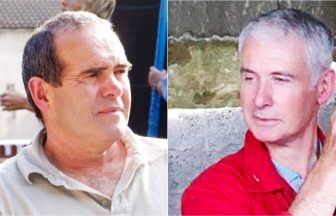
Professor Marcel Schubert from the University of Cologne explained: “We developed microlasers that are injected directly into embryos or mixed into artificial tumours.
“The microlasers are actually droplets of oil that are doped with a fluorescent dye.
“As the biological forces get to work, the microlasers are squished and deformed by the cells around them.
“The laser light changes its colour in response and reveals the force that’s acting upon it.
“In real time we could measure and monitor biological forces.”
 PA Media
The researchers say the oil and dye used to make the microlasers are non-toxic and made from readily available materials, so they don’t interfere with biological processes and could become a commercially viable technology.
Professor Malte Gather, University of St Andrews, said: “Embryos and tumours both start with just a few cells.
“It is still very challenging to understand how they expand, contract, squeeze and fold as they develop.
“Being able to measure biological forces in real time could be transformative.
“It could hold the key to understanding the exact mechanics behind how embryos develop, whether successfully or unsuccessfully, and how cancer grows.”
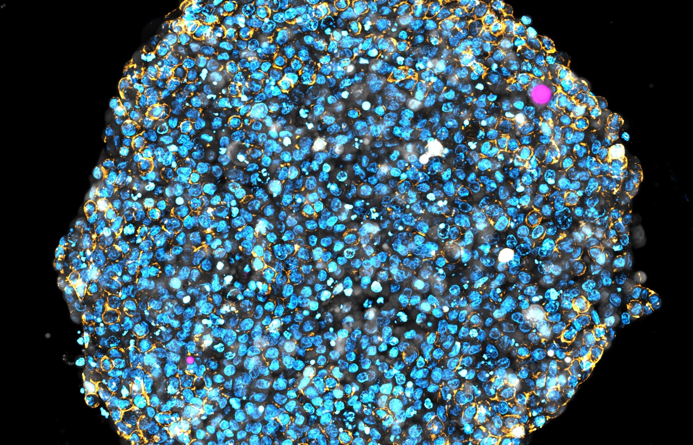
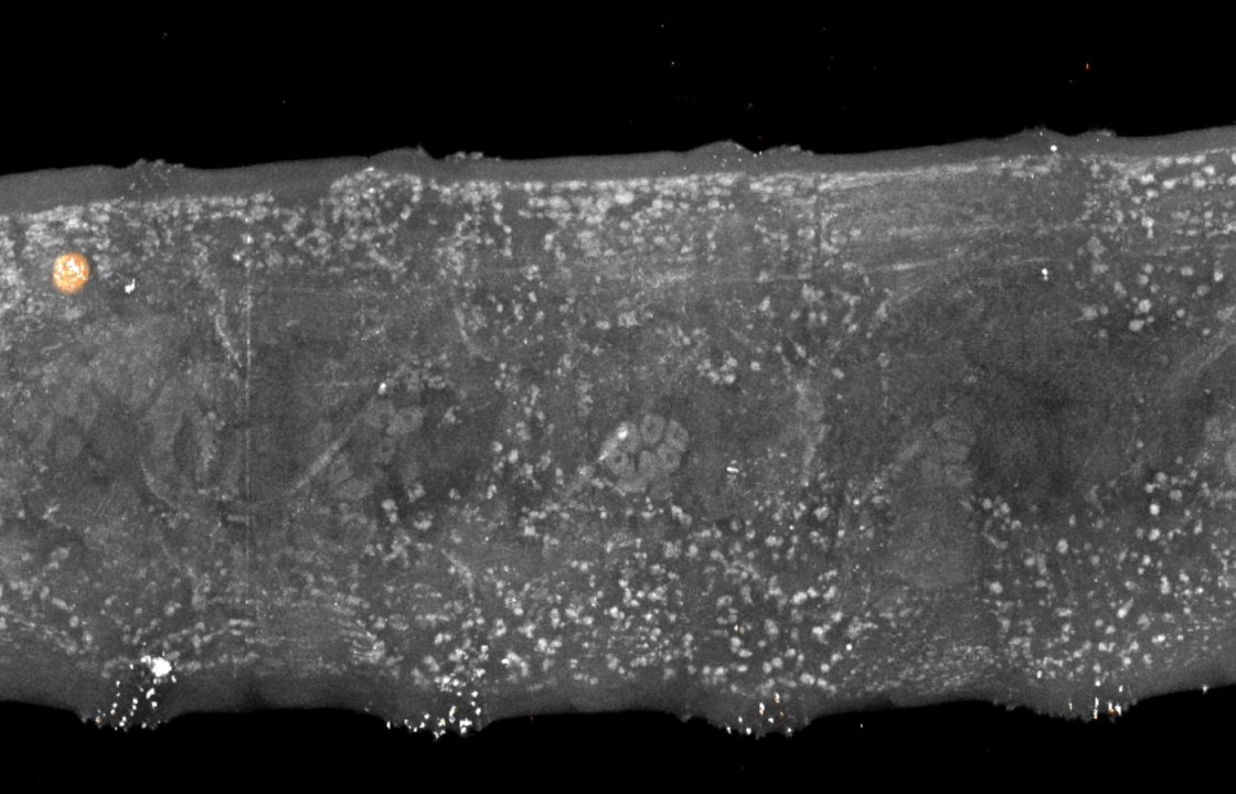
The researchers tested their method on fruit fly larvae and in “tumour spheroids” – artificial tumours made from brain tumour cells.
Professor Gather explained: “We measured the 3D distribution of forces within tumour spheroids and made high-resolution long-term force measurements within the fruit fly larvae.”
The team now hopes hope to gain funding to adapt it for clinical trials, adapting the method as required for larger cell systems.
The study, which is published in the journal Light: Science & Applications, was funded by a number of bodies including the Engineering and Physical Sciences Research Council, the Humboldt Foundation and the European Union’s Horizon 2020 Framework Programme.
Follow STV News on WhatsApp
Scan the QR code on your mobile device for all the latest news from around the country


 PA Media


